Sunday, 18 December 2016

REPRODUCTIVE HEALTH


CHAPTER 4
REPRODUCTIVE HEALTH

WHO - WORLD HEALTH ORGANISATION definition
Health: Health is a total well-being in all aspects of reproduction, i.e., physical, emotional, behavioral & social; and not merely the absence of disease or infirmity.

Reproductive health is defined as a state of physical, mental, and social well-being in all matters relating to the reproductive system, at all stages of life; and not merely the absence of disease or infirmity.
Reproductive health addresses the reproductive processes, functions and system at all stages of life. It, therefore, implies that people are able to have a responsible, satisfying and safe sex life and that they have the capability to reproduce and the freedom to decide if, when and how often to do so.

Reproductive Health; Importance:
Reproductive health is a crucial part of general health and a central feature of human development.  It is a reflection of health during childhood, and crucial during adolescence and adulthood, sets the stage for health beyond the reproductive years for both women and men, and affects the health of the next generation.  The health of the newborn is largely a function of the mother's health and nutrition status and of her access to health care.
Factors affecting reproductive health
Reproductive Health– problems and strategy:

India was among the 1st countries to initiate actions & plans to attain total reproductive health as social goal.
These programmes are called as ‘FAMILY PLANNING’
Improved programmes currently in operation have a popular name ‘Reproductive & Child Health Care Programmes’ (RCH). Launched in 1977.

Sexual and Reproductive Health Awareness Day is held annually on February 12.

Problems:
·        Lack of awareness and readiness to discuss sex related issues
·        Myths and misconceptions related to sex
·        Early marriages and lack of prenatal and post natal care, leading to high maternal mortality
·        Unwanted or mistimed pregnancies
·        Prevalence of sexually transmitted diseases and reproductive tract infections
·        Population explosion
·        Illegal abortion of female fetuses
·        Congenital or acquired infertility

Aims of Reproductive Health Programme

·        To create awareness about various aspects of and related to reproduction
·        To educate adolescents regarding the significance of reproductive health, making the right choices and providing a healthy understanding of the sex related issues.
·        To lower the incidences of sexually transmitted diseases, and reproductive tract infections, by creating awareness regarding the significance of safe and hygienic sexual practices and proper care of reproductive organs.
·        To address issues of fertility, ensure counseling and guidance to couples incapable of bearing children. Development of assisted reproductive technologies (ARTs).
·        To educate regarding the importance of prenatal, and post natal care of mother and child.
·        To make people aware of legal issues related to reproductive process and technologies such as female foeticide and amniocentesis.
·        To develop technology for faster dissemination of knowledge and use of audio visual methods for developing a better understanding.

HOW HAS THE GOVERNMENT TAKEN MEASURES?

·        The use of audio-visuals & print media to create awareness
·        Parents, family members, close relations, teachers and friends are involved in the awareness programmes.
·        Introduction of Sex education in schools. Young students are most vulnerable to misconceptions and hence it was considered important to introduce sex education to them.
·        Proper information about reproductive organs, adolescence and related physiological changes, safe & hygienic sexual practices, sexually transmitted diseases, AIDS etc. 
·        Educating fertile couples, especially those of marriageable age group about available birth control options, proper care of pregnant mothers, pre and post natal care, importance of breast feeding etc.
·        Development of understanding of male and female physiology, their respective social and biological roles an in the light of this information; development of gender equality.
·        Awareness of problems related to population explosion, both at family and society level. Importance of small family, and limited resources.
·        Awareness of social evils and sex related crimes. Educating children to identify, and understand as well as protect themselves from these evils.
·        Development of infrastructure, technology and medical expertise to help people in need.
·        Research on reproduction related areas, to develop new method and improve existing methods.
·        Statutory ban on amniocentesis, massive child immunization programme etc.
·        Legalisation of MTP (medical termination of pregnancy) in 1971, to control population size by check unwanted pregnancies.
Overall development goal of measures taken by government is building up of a socially responsible and healthy society.

Amniocentesis
A fetal sex determination test based on the chromosomal pattern in the amniotic fluid surrounding the developing embryo.
It is used to find out chromosomal abnormalities in developing embryo by using amniotic fluid.
It is also illegally used for foetal sex determination and hence a statutory ban on amniocentesis has been placed to check female foeticide.

Indicators of Reproductive Health
·        Awareness of sex and reproduction related matters
·        Higher number of medically assisted deliveries
·        Better pre and post natal care, indicated by lower maternal and infant mortality
·        Smaller families with more couples opting for family planning programmes
·        Early detection and timely cure of STDs and other sex related problems
·        Higher number of ARTs, reducing problems of infertility

Population explosion & Birth control:
Scientific study of human population: Demography

 World population (1900): 2 billion                    India’s Population (1947): 350 million
World Population (2001): 6 billion                                India’s Population (2000): 1b

v  India is the second most populous country in the world
v  India’s population reached 1 billion on 11 may, 2000

Population Index:
Ø  Birth Rate (Natality rate): Natality rate is the rate at which new individuals are added to a particular population by reproduction (birth of young ones or hatching of eggs or germination of seeds/spores). It is generally expressed as number of births per 1,000 individuals of a population per year.
Ø  Death rate (Mortality rate): Mortality rate is the rate at which the individuals die or get killed. It is the opposite of natality rate. Mortality rate is generally expressed as number of deaths per 1,000 individuals of a population per year.
Ø  Vital Index: The percentage ratio of natality over mortality expressed in percentage is called Vital index.
Vital index determines the normal rate of growth of a population.

Ø  Growth Rate: The "population growth rate" is the rate at which the number of individuals in a population increases in a given time period. It is expressed as a fraction of the initial population.

Ø  Total fertility rate (TFR): TFR is defined as the average number of children that would be born to a woman in her lifetime if the age specific birth rates remain constant. The value of TFR varies from 1.9 in developed nations to 4.7 in developing nations. 


Causes of Population Explosion
·        Increased food production and distribution
·        Improvement in public health due to better sanitation, and cleaner water
·        Improvement in medical technology leading to conquest of diseases though early detection, better health practices, new medicines, vaccinations etc.
These factors have resulted in:
·        Decline in death rate
·        Decline in MMR (maternal mortality rate)
·        Decline in IMR (infant mortality rate)
·        Increase in number of people in reproductive age


According to the Census of 2001- 2011, India's population has grown at 17.7 per cent as against 21.5 per cent in the decade. At this rate it will double in next 33 years. This would lead to scarcity of basic requirements for life; food, shelter and clothing. Hence the government has been forced to take significant measures for population control.

Methods to Control population:
Ø  Education
Ø  Raising age of marriage
Ø  Family Planning methods

Steps Taken to execute the above:
·        Targeting an increase in literacy rate
·        Involving social organizations to make family planning programme a people’s programme
·        Providing education and employment opportunities to women
·        Proper implementation of community health programme
·        Incentives to people undergoing sterilization
·        Providing birth control facilities to individuals

CONTRACEPTIVE METHODS:

Through media – HUM DO HAMARE DO!!!!! (WE 2, OUR 2)
SAHELI - it is a contraceptive method developed by scientists in CDRI - Central Drug Research Institute.

Contraceptive Methods:

Natural Methods
Barriers
 I U D
Oral contraceptives
Injectable implants
Surgical Methods

1. Natural Methods:
Avoids  meeting of sperm & ovum.

Periodic Abstinence - Avoid coitus from day 10 – 17 of  menstrual cycle when ovulation is expected. Because chances of fertility is very high during this period, hence known as fertile period.
Withdrawal or coitus interuptus - Male partner withdraws his penis from vagina before ejaculation avoiding insemination of sperms
Lactational amenorrhea-Absence of menstrual cycle during first six months of intense lactational period.

2. Barrier Methods:

Condoms - Thin rubber used to cover penis in male or vagina & cervix in females.
Diaphragms, Cervical caps & vaults are all barriers for females to cover cervix during coitus.

Advantages of barrier methods:
1.       They are disposable.
2.       They can be self –inserted.
3.       They are reusable.
4.       Prevents conception by blocking entry of sperm thru cervix.
3. Intra uterine  devices (IUD’S):

Devices   inserted by doctors or nurses in uterus thru vagina.
Examples. Cu T, Cu7, Multiload 375, Lippes loop.

Cu ions released suppress sperm motility & fertilizing capacity of sperms.
Hormone releasing IUDs makes the uterus unsuitable for implantation & cervix hostile to the sperm.

4. Oral pills:

Pills are taken daily for 21 days.
They are very effective with less side effects.
Saheli - new oral contraceptive contains a non-steroidal preparation.
It is a ‘once a week ‘pill with high contraceptive value.
Injection or implantation of progesterone /estrogen under the skin.

5. Surgical  method:

This method is also called as STERILISATION.
It is advisable for male/female partner as a terminal method to prevent any more pregnancies.
In male, they’re called vasectomy, where the vas deferens is cut or tied.
In female, it is called tubectomy, where a small part of the fallopian tube is cut or tied up.
This method is highly effective but their reversibility is very poor.

Side effects of contraceptive method:
•       It is very important that the selection of contraceptive method should be taken under the consultation of the doctors.
•       However, their possible ill-effects like nausea, abdominal pain, breakthrough bleeding, irregular menstrual bleeding or even breast cancer.
•       These symptoms SHOULD NOT BE TOTALLY IGNORED.

What is MTP?
Intentional or voluntary termination of pregnancy before full term is called Medical Termination of Pregnancy (MTP) or Induced Abortion.

Why MTP?

MTP is done to get rid of unwanted pregnancies due to casual unprotected intercourse or failure of the contraceptive used during coitus or rape.
MTPs are also essential in certain cases where continuation in pregnancy could be harmful or even fatal to the mother or to the foetus or both.

Sexually transmitted diseases ( STDs)
Diseases or infections which are transmitted sexually through sexual intercourse are called as Sexually Transmitted Diseases (STDs) or Venereal Diseases (VDs) or reproductive tract infections. STDs can be classified as viral, bacterial, protozoan, fungal, etc.

HOW ARE STDS CAUSED?
Depending on the disease, STDs can be spread with any type of sexual activity. STDs are most often caused by viruses and bacteria.

Various types of Sexually Transmitted Diseases:
The various types of sexually transmitted diseases include gonorrhoea, syphilis, genital herpes, chancroid and of course the most common HIV leading to AIDS.

Chlamydiasis

Chlamydiasis is a sexually transmitted disease in humans caused by the bacterium Chlamydia trachomatis. Chlamydiasis is a major infectious cause of human genetial and eye diseases.
Chlamydiasis was once the most important cause of blindness. the infection can spread from eye to eye by fingers, shared towels, eye seeking flies, and cloths etc.

Prevention
STDs are a major threat to a healthy society. Therefore early detection or prevention and cure of these Diseases are given prime consideration under Reproductive health-care programmes though all person are vulnerable to these infections, their incidences are reported to be very high among the age group of 15-24years.these infections can be prevented by following a few simple rules which include:

Avoid sex with unknown partners or multiple partners
Always use condoms during coitus
In case of doubt, go to a qualified doctor for early detection and get complete treatment if diagnosed with disease.

INFERTILITY
A large no of couples all over India are infertile, i.e., they are unable to produce children in spite of Unprotected sexual co-habitation. The reasons for this could be many-physical, congenital, diseases, drugs, Immunological or even Psychological.

Assisted Reproductive Technologies (ART) are special techniques that assist couples to have children.
VARIOUS TYPES OF ASSISTED REPRODUCTIVE TECHNOLOGIES ( ART)INCLUDE:
·         In-vitro fertilisation (IVF)
·         Zygote intra fallopian transfer (ZIFT)
·         Intra cytoplasmic sperm injection(ICSI)
·         Gamete intra fallopian transfer(GIFT)
·         Artifical insemination (AI)

1) In Vitro Fertilization (IVF)
It is the fertilization outside the body in almost similar conditions as that in the body. In this method, popularly known as test tube baby programme, ova from the wife / donor (female) and sperms from the husband / donor (male) are collected and are induced to form the zygote under simulated conditions in the lab. The zygote or early embryos could then be transferred into the fallopian tube (ZIFT -zygote intra fallopian transfer)

2) Zygote intra fallopian transfer (ZIFT)
The zygote with 8 blastomeres can be transferred into the fallopian tube.

3) Intra cytoplasmic sperm injection (ICSI)
Intra cytoplasmic sperm injection (ICSI) is another specialized procedure to form an embryo in the lab in which a sperm is directly injected into the ovum.

4) Gamete intra fallopian tube (GIFT)
Transfer of an ovum collected from a donor into the fallopian tube of another female who cannot produce one, but can provide suitable environment for fertilisation and further development is another method attempted.

5) Artificial Insemination (AI)
Infertility cases either due to inability of the male partner to inseminate the female or due to very low sperms counts in the ejaculates could be corrected by artificial insemination.

In the technique, the semen collected either from the husband or a healthy donor is artificially introduced into the vagina or into the uterus (IUI - Intra Uterine Insemination) of the female.

6) Adoption – can be done from orphanage / relatives.

Counseling and information on infertility

It is important to involve both partners in all aspects of management. Discussions of wishes, plans, beliefs and motives are important.
Tuesday, 14 July 2015

HUMAN REPRODUCTION

Chap 3

HUMAN REPRODUCTION

Human reproductive system: organ system by which humans reproduce and bear live offspring
Provided all organs are present, normally constructed, and functioning properly, the essential features of human reproduction are:
  1. liberation of an ovum, or egg, at a specific time in the reproductive cycle
  2. internal fertilization of the ovum by spermatozoa, or sperm cells
  3. transport of the fertilized ovum to the uterus, or womb
  4. implantation of the blastocyst
  5. the early embryo developed from the fertilized ovum, in the wall of the uterus
  6. formation of a placenta and maintenance of the unborn child during the entire period of gestation
  7. birth of the child and expulsion of the placenta, and
  8. Suckling and care of the child, with an eventual return of the maternal organs to virtually their original state.

Human beings exhibit sexual reproduction and vivipary.
Like all mammals human beings are unisexual with either the male or female reproductive system.
Of the reproductive structures, the gonads (testes in males and ovaries in females) are considered as the primary sex organs, while other associated structures including reproductive ducts and glands are known as secondary sex organs.

Table 1: Secondary sex organs
Male
Epididymis, vasa deferens, prostrate, seminal vesicles, penis
Female
Fallopian tubes, uterus, vagina, mammary glands

Human beings also exhibit sexual dimorphism, i.e. male and female exhibit certain specific morphological features make them distinct from the other sex. These characters are known as secondary or accessory sexual characters, and the phenomenon is known as Sexual Dimorphism.

Basic Phases of reproductive physiology:
The reproductive events in humans include:
  1. Gametogenesis: Formation of gametes.
Male gametes or Sperms and female gametes or ova (singular ovum), are haploid sex cells formed from diploid germ cells. The germ cells are present in primary reproductive organs called gonads. Gonads in male are testes and in females are ovaries.
Gametogenesis in males involves formation of sperms – Spermatogenesis
Gametogenesis in females involves formation of ova – Oogenesis

  1. Insemination: Transfer of sperms in to female genital tract
  2. Fertilization: Fusion of male and female gametes to form diploid zygote
  3. Implantation: Involves 2 steps. First, rapid cell division of zygote leads to formation of single layered hollow spherical larva called blastula. The process is known as blastulation.


Next the blastula becomes attached to the endometrium of uterine wall. This step is known as implantation.
  1. Gestation: Embryonic development. It involves:
    • Placentation: involves the formation of an intimate physiological and mechanical connection between the foetal and maternal tissues to enable nutrition, respiration, and excretion etc of the fetal tissue.
    • Gastrulation: Formation of gastrula larva from the blastocyst. Gastrula larva has three primary germ layers.
    • Oganogenesis: Formation of specific organ system from the three germ layers.

In humans the gestation period is of 280 days.
  1. Parturition: Child delivery of the fully formed human baby.

MALE REPRODUCTIVE SYSTEM


Located in pelvis region
Comprises of:
  • A pair of testes
  • Accessory ducts
  • Glands and
  • External genitalia

                   I.      Testes: Primary sex organ.1 pair.
Oval, 4-5cm in length, 2-3 cm in width
Situated below the abdominal cavity within a pouch known as scrotum or scrotal sacs
The scrotal sacs are filled with a tissue fluid called hydrocoel. Gubernaculum and spermatic cord keep the testes in position inside the scrotal sacs.
Scrotal sacs help in maintaining the low temperature of the testes (at 2-2.5 deg C lower than the normal internal body temperature. This is important for normal; spermatogenesis since high body temperature kills spermatogenic tissue.

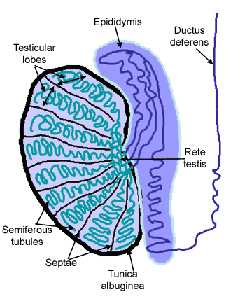
Each testis is covered by a dense white fibrous capsule known as tunica albuginea.  It projects inside the testes as fibrous septa. The septa divide the testis into 250 compartments called testicular lobules.

Each of the testicular lobule has 1-3 highly coiled seminiferous tubules. The seminiferous tubules produce sperms. The seminiferous tubules are lined on the inside by two types of cells:
·        Male germ cells or Spermatogonia: produce sperms
·        Sertoli or nurse cells: Provide nutrition to germ cells
The germ cells undergo meiotic divisions and form haploid and motile gametes known as spermatozoa.


The region in between the seminiferous tubules is known as interstitial spaces. It contains small blood vessels and interstitial cells or leydig cells. Leydig cells synthesize and secrete testicular hormone called androgens. The most important androgen is testosterone, this controls the development of secondary sexual characters and spermatogenesis.
Function of testes: spermatogenesis and secretion of testosterone

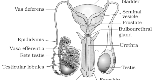
                            II.      Accessory ducts: the male sex accessory ducts include
a.      Rete Testis
b.      Vasa efferentia
c.       Epididymis
d.      Vasa deferentia
Seminiferous tubules open into a network of tubules known as rete testis which further leads in to vasa efferentia.
The vasa efferentia leaves the testes and opens in to epididymis on the posterior surface of each testis.
The main function of epididymis is conduction of sperms by peristalsis. Epididymis also helps in storage nutrition and physiological maturation of sperms by removing certain decapacitation factors.
Epididymis leads to vasa deferens that ascends to the abdomen and loops over the urinary bladder. It receives a duct from the seminal vesicle and opens in to urethra as the ejaculatory duct.
                         III.      Urethra: arises from the urinary bladder, joins the ejaculatory duct to form the urinogenital tract. It extends through the penis. This tract carries urine, sperms and secretions of seminal vesicles, Cowper’s glands.
                          IV.      Penis: male external genitalia
Made of special tissue that helps in erection of the penis to facilitate insemination. Tip of penis is highly sensitive and enlarged. It is called glans penis. The tip of glans penis is covered by a loose retractile skin known as foreskin. Glans also has a slit like opening of urinogenital tract called urethra maetus.
                             V.      Male Accessory Gland: Include
·        Seminal vesicle: secretions comprise 60-70% of semen. Mainly formed of fructose, citrate, several proteins and prostaglandins. Activate spermatozoa and stimulate vaginal contractions to help in fusion of gametes.
·        A Prostrate gland: Surrounds the proximal part of urethra. Pours alkaline secretions in urethra. Secretions contain lipids, small amount of citric acid, bicarbonate ions and some enzymes e.g. fibrinolysin.
It forms 20% of semen. Main functions are:
o       Activates sperms
o       Provides nutrition to sperms
o       Neutralizes acidity of urine
o       Adjusts vaginal pH
·        A pair of bulbourethral glands: secretes mucus like alkaline substance which provides lubrication to penis.

Secretions of these glands constitute the seminal plasma which is rich in fructose, calcium and certain other enzymes.
Thus the major functions of male reproductive system include:
ü      Spermatogenesis
ü      Secretion of male hormone testosterone
ü      Transfer of sperms to female reproductive system for fertilization of ovum

FEMALE REPRODCUTIVE SYSTEM
FIGURE 3.3

Located in pelvis region
Comprises of:
·        A pair of ovaries
·        A pair of oviducts
·        uterus
·        cervix
·        vagina
·        external genitalia
Fallopian tubes, uterus and vagina collectively form the accessory ducts.
The above organs along with mammary glands are together involved in the reproduction which includes the following processes:
o       ovulation
o       fertilization
o       pregnancy
o       birth and
o       child care

1.      Ovaries: Primary female sex organs, produce ovum and several steroid hormones (ovarian hormones)

o       1 pair, small sized (2-4 cm), almond shaped
o       present in pelvis region, one on each side of uterus
o       suspended from abdominal wall by mesovarium
o       attached to pelvic wall and uterus by ovarian ligament
Each ovary is covered by a thin germinal epithelium. The epithelium surrounds the stroma that is divided in to 2 zones:
  • Outer Cortex
  • Inner medulla
In the medulla of ovaries are present a number of developing or primary ovarian follicles in different stages of oogenesis.
Cortex may contain either a yellow conical endocrine gland known as corpus luteum or a degenerating white corpus albicans.
2.      Fallopian tubes or Oviducts: each is 10-12 cm long, and extends from periphery of each ovary to the uterus.
The fallopian tubes comprise of 3 parts:
a. Infundibulum: Part closer to ovary is funnel shaped infundibulum. The ends of infundibulum have finger like projections known as fimbriae. Fimbriae help in collection of ovum after ovulation.
b. Ampulla: The infundibulum leads to a wider ampulla.
c. Isthumus: The ampulla leads the last part of oviduct which has a narrow lumen and leads in to uterus. It is ciliated.
Ampullary isthmic junction is the site of fertilization
3.      Uterus or Womb: single, inverted pear shape, muscular and highly vascular. Supported by ligaments attached to the pelvic wall.
Formed of three parts:
·        Fundus: upper dense part
·        Body or corpus: middle and main part. The wall of uterus in this region is made of three layers.

Table 2: Uterine wall layers and their functions
Perimetrium
External, thin and membranous
Forms envelope
Myometrium
Middle thick layers of smooth muscles
Helps in child birth by undergoing strong contractions
Endometrium
Inner and Glandular layer
Undergoes cyclical changes during menstrual cycle
Site of implantation and foetal growth during pregnancy
Sit of formation of placenta

·        Cervix: lower narrow part. Uterus opens in to vagina through cervix. Cavity of cervix known as cervical canal, which along with vagina forms the birth canal.
4.      Vulva: external genitalia of the female. It comprises of:
·        Vestibule or a depression that has two apertures: upper urethral orifice and a lower vaginal orifice. The vaginal orifice is partially covered by a membranous fold called hymen which is torn either during the first coitus, or due to an incident of sudden fall, jolt, physical activity such as sports, horse riding, cycling, or during insertion of tampons. In some women hymen may persist even after coitus.
·        Vestibule is bounded by two pairs of moist fleshy skin folds with sebaceous glands: inner smaller labia minora and outer larger labia majora.
·        The labia minora fuse anteriorly to form a skin fold known as prepuce which lies in front of a small erectile organ glans clitoris (homologous to penis).
·        A fleshy elevation is present above labia majora, known as mons pubis. It is covered by skin and pubic hair.

5.      Mammary Gland: A functional mammary gland is a characteristic feature of mammals.
Mammary glands are paired structures, containing glandular tissue and fats.
The glandular tissue is divided in to 15-20 mammary lobes containing clusters of cells known as alveoli. The cells of alveoli secrete milk, which is stored in the lumen of alveoli.
The alveoli open into mammary tubules. The mammary tubules join to collectively form mammary duct. Several mammary ducts join to form wider mammary ampulla. The mammary ampulla is connected to lactiferous duct through which milk is sucked out.

The uterus, oviducts, vagina, clitoris, the accessory genital glands and the mammary glands are referred to as secondary sex organs of female.

Major Functions of Female Reproductive System
  1. Oogenesis by germ cells of ovary
  2. Receiving seminal fluid
  3. Fertilization of gametes to form zygote
  4. Implantation followed by prenatal growth
  5. Parturition
  6. Post natal care

Gametogenesis
Process of formation and differentiation of haploid gametes (sperms and ova), from diploid primary germ cells (gametogonia)
The gametogonia (spermatogonia and oogonia) are present in primary sex organs or gonads; testes in male and ovaries in female.
Thus gametogenesis can be of 2 types:
  1. Spermatogenesis
  2. Oogenesis

Spermatogenesis
(fig 3.5)

Formation of haploid and functional male gametes (spermatozoa, n) from diploid reproductive cells, spermatogonia (2n) present in testes of male organism.
Occurs in testes
Begins at puberty
Continuous process completed in 74 days
 Process: Divided in to 2 phases:
  1. Formation of Spermatids:
  • Multiplicative or mitotic phase: The spermatogonia (2n) present on the inside wall of seminiferous tubules multiply by mitotic division and increase in number. Each spermatogonium is diploid and contains 46 chromosomes.
  • Growth Phase: Diploid
  • Some of these spermatogonia called primary spermatocyte periodically undergo meiosis. A primary spermatocyte completes first meiotic division (reduction division) leading to formation of two equal haploid cells called secondary spermatocyte, which have only 23 chromosomes.
  • The secondary spermatocytes (n) undergo second meiotic division to produce four equal haploid spermatids (n).
  • Spermatids are non-motile.

  1. Spermiogenesis
                     i.            The spermatids are transformed into spermatozoa (sperms, n) by the process called spermiogenesis. Spermiogenesis converts the non motile spermatids in to functional and motile spermatozoa.
The main aim of spermiogenesis is to increase sperm motility by reducing weight and development of locomotory structure.
                   ii.            After spermiogenesis, sperm heads become embedded in sertoli cells which provide nourishment to them.
                  iii.            Mature spermatozoa are finally released from seminiferous tubules by the process called spermiation.

Sperms once released from seminiferous tubules, are transported by accessory ducts. Secretion of epididymis, vas deferens, seminal vesicle, and prostrate are essential for maturation and motility of sperms. Thus:
Sperms+Seminal Plasma=Semen
The functions of male sex accessory ducts and glands are maintained by the testicular hormones (androgens).

Structure of Sperm
(figure 3.6)

Ø      Microscopic
Ø      Composed of Head, Neck, Middle piece and a Tail
Ø      A plasma membrane envelops the whole body
Ø      Head contains an elongated haploid nucleus. Its anterior part is covered by a cap like structure called acrosome.
Acrososme is formed from a part of the Golgi Body of the spermatid. It is filled with enzymes that help fertilization of the ovum. The lytic enzyme hyaluronidase present in acrosome is responsible for penetration of ovum during sperm entry.
Ø      The middle piece has many mitochondria. They produce energy required for the movement of tail that causes sperm motility. Sperm motility is essential for fertilization.
Ø      Tail is the longest part of sperm. It exhibits lashing movements that provide forward push to the sperm.
A human male ejaculates about 200 to 300 million sperms during coitus of which for normal fertility at least 60% sperms must have normal shape and size; and at least 40% must have normal motility.
Viability of human sperm is 24 hours.

Control
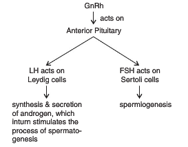
Spermatogenesis starts at the age of puberty due to significant increase in secretion of gonadotrophin releasing hormone (GnRH). It is a hypothalamic hormone.
High level of GnRH acts on anterior pituitary and stimulates release of 2 gonadotrophins:
  • Leutinising Hormone (LH): Acts at Leydig cells and stimulates synthesis and secretion of androgens.
Androgens stimulate process of spermatogenesis
  • Follicle Stimulating Hormone (FSH): acts on Sertoli cells, and stimulates secretion of some factors which help in process of spermiogenesis.
Control of Spermatogenesis


Oogenesis:
Process of formation of natural haploid female gamete called ova (n) (singular: ovum), from diploid egg mother cells Oogonia (2n) of ovary.
Initiated during embryonic development stage. A couple of million gamete mother cells are formed within each foetal ovary. No more are formed and added after birth.
The process involves three phases:
  1. Multiplicative Phase: Certain primary germ cells of the germinal epithelium of ovary undergo rapid mitotic divisions to form groups of diploid egg mother cells or oogonia (2n).
  2. Growth Phase: The oogonium start division and enter prophase I of meiosis. The division gets arrested at that stage forming primary oocyte.

    • Each primary oocyte then gets surrounded by layer of granulosa cell and is then called Primary Follicle. Many of these primary follicles degenerate during the phase from birth to puberty. By puberty only about 60,000 – 80,000 primary follicles remain in each ovary.
    • Each Primary follicle then gets surrounded by more layers of granulosa and a theca forming the secondary follicle.
    • Secondary follicle than develops a fluid filled antral cavity called antrum and is now termed as Tertiary Follicle. The theca layer gets organized in to inner theca interna and outer theca externa.

  1. Maturation Phase:
    • At this stage the primary oocyte within the tertiary follicle grows in size and completes its first meiotic division. It is an unequal division resulting in formation of a large secondary oocyte and a tiny first Polar Body.
    • The secondary oocyte retains bulk of the nutrient rich cytoplasm of the primary oocyte.
    •  The tertiary follicle further changes into Graffian Follicle.
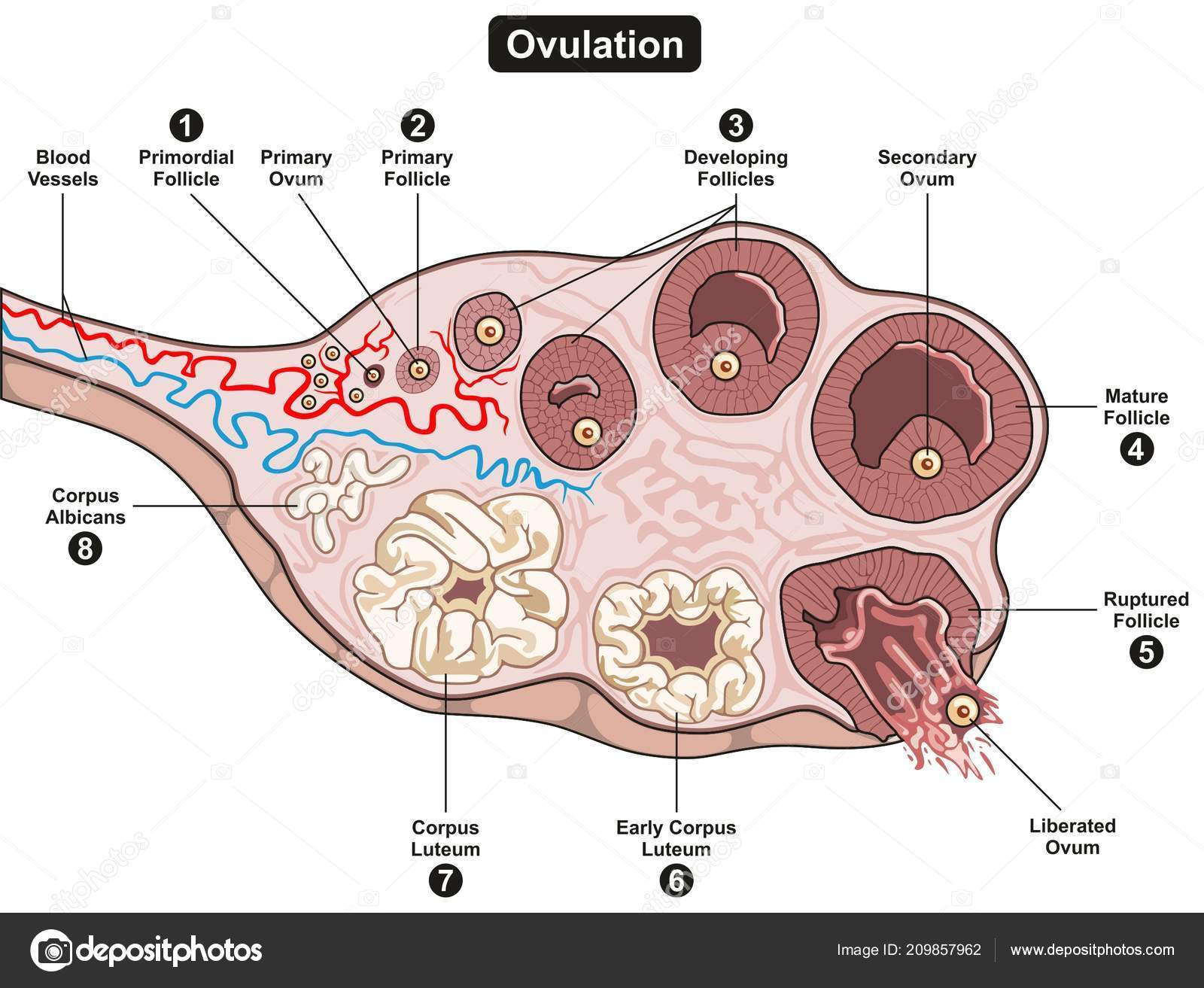
    • The secondary oocyte forms a new membrane called Zona Pellucida surrounding it. The Graffian Follicle now ruptures to release the secondary oocyte (Ovum) from the ovary by the process called Ovulation.

Structure of Ovum

  • Maternal haploid gamete
  • Generally spherical, non motile with yolky cytoplasm enveloped in 1 or more egg envelopes
  • Human ovum is alecithal with negligible amount of cytoplasm
  • Cytoplasm is differentiated in to outer, smaller and transparent exoplasm or egg cortex; and inner larger and opaque endoplasm or ooplasm
  • Nucleus is large and excentric
  • The side of ovum with nucleus and polar body is known as Animal Pole; and the opposite side is known as Vegetal Pole.
  • Ovum is surrounded by many egg envelopes:
  • Vitelline Membrane: inner, thin and transparent
  • Zona Pellucida: middle, thick and transparent
  • Corona Radiata: outer and thick


Spermatogenesis vs. Oogenesis


Menstrual Cycle:

The cyclic changes that occur in the reproductive organs of primate females is called menstrual cycle.
Menarche: The beginning of first menstruation at puberty.
Menopause: Menstrual cycle cease at the age of 45 – 50. This is known as menopause.
In human females the menstruation is repeated at an average interval of 28/29 days. The entire cycle starting from one menstruation to next, is known as menstrual cycle. The cycle involves the release of an ovum during the middle of the cycle.

There are typically 4 phases in menstrual cycle, controlled by two types of hormones: follicle stimulating hormones (FSH) and luteinising hormones (LH). 
Menstrual phase
It lasts for 3-5 days
It results due to breakdown of endometrial lining of uterus and its blood vessels
Menstruation occurs only if fertilization of the ovule does not take place. Lack of menstruation may be indicative of ovule fertilization and hence of pregnancy. However, it may also be due to stress, poor diet, poor health etc.
Follicular phase
Primary follicle in the ovary grow to become a fully mature Graffian follicle. Simultaneously, Endometrium is regenerated by proliferation of its cells
These changes are due to increased levels of pituitary and ovarian hormones: Gonadotropins (FSH, LH), Estrogen 
FSH controls follicular phase, stimulates growth of follicles.
Secretion of FSH and LH, increases gradually during the follicular phase. It performs 2 functions:
  • Stimualtes follicular development
  • Stimulates secretion of estrogen by growing follicles
Secretion of Estrogen occurs during the follicular phase.
Ovulatory phase
Involves ovulation. Ovulation of mature follicles on the ovary is induced by a large burst of LH secretion known as the preovulatory LH surge
Peak level of LH induces rupture of mature Graafian follicle and release of ovum or ovulation.
Luteal phase
Ruptured follicle transforms into corpus luteum 
Residual cells within ovulated follicles proliferate to form corpora luteum, which secrete the steroid hormones progesterone and estradiol. Progesterone is necessary for maintenance of pregnancy by maintaining endometrium. In most mammals, LH is required for continued development and function of corpora luteum. [The name luteinizing hormone derives from this effect of inducing luteinization of ovarian follicles.]
During pregnancy, all events of all events of menstrual cycle stop and there is no menstruation.
In absence of fertilisation, corpus luteum degenerates. This causes disintegration of endometrium leading to menstruation, marking the beginning of a new cycle.

Fertilisation and Implantation

The process of fusion of a sperm with an ovum is called fertilisation.
Fertilisation can only occur if the ovum and sperms are transported simultaneously to the ampullary – isthmic junction.

Events during fertilisation:

  1. 200-300 million sperms are introduced in to female genital tract.
    • About 50% of these are killed due to acidity in the female genital tract.
    • Many are engulfed by the phagocytes of the vaginal epithelium.

Thus around 100 sperms only reach the fallopian tubes.
  1. Ovulation or release of ovum from graffian follicle of the ovary occurs on the 14th day. At the time of ovulation, the ovule is at secondary oocyte stage.
  2. Penetration of Sperms: Sperm comes in contact with egg at animal pole. Sperm penetration is a chemical phenomenon. Acrosome of sperm comes in contact with zona pellucida. Acrosome releases certain sperm lysins that dissolves egg envelope locally and thus make path for penetration of sperm. The lysing enzyme present in sperm lysins is Hyaluronidase. It penetrates through corona radiate and dissolves zona pellucida.
Only the sperm nucleus and middle piece enter the ovum, the tail is lost.

  1. In humans there is always monospermy. Entry of sperm induces changes in membrane that block entry of additional sperms

  1. Entry of sperm induces:

  • Meiotic division of secondary oocyte to form haploid ovum and 2nd polar body.
  • Formation of zygote

Significance of fertilization:
                     i.            Stimulates the secondary oocyte to undergo second maturation division to release second polar body and to form haploid ovum.
                   ii.            Restores diploidy
                  iii.            Fertilization membrane prevents polyspermy
                 iv.            Combines characters of two parents and introduces variations. So helps in evolution
                   v.            Centrioles of sperm from the spindle to initiate the cleavage of zygote


Cleavage
Rapid mitotic division of zygote to from hollow, spherical multicellular developmental stage called blastula.. So the process is also called bl;astulation.
Ø      Formation of Morula
  • In human zygote cleavage occurs in fallopian tubes during the passage of zygote towards the uterus.
  • It is holoblastic[p1] , radial, indeterminate and unequal.
  • The zygote divides first in to 2 unequal cells called blastomeres.
  • Second cleavage is perpendicular to first.
  • Subsequent divisions occur rapidly one after the other leading to formation of Morula.
  • Morula is 16-32 cell stage
Ø      Formation of Blastula
·        Rearrangement of Blastomeres
·        Outer layer of cells becomes flat and forms trophoblast, that draws nutrition from endometrium of uterus
·        The inner cell mass is known as micromeres.
·        The central cavity is known as blastocoel, in which the nutrition collects.
Implantation
Process of attachment of blastocyst on the endometrium of uterus
Blastocyst gets embedded in endometrium of uterus (implantation)
Uterine cells rapidly divide and cover the blastocyst 
Implantation may occur anytime between 6th and 10th day after fertilization (usually 7th day)

Pregnancy and embryonic development

Inner layer grows out as finger like projections called villi into the uterine stroma
Chorionic villi and uterine tissue get interdigitated to form placenta
Placenta secretes hormones like hCG , hPL , estrogens , progesterones (to maintain pregnancy)
Inner cell mass differentiates into an outer layer called ectoderm and an inner layer called endoderm
Mesoderm appears between ectoderm and endoderm
Stem cells (undifferentiated embryonic cells)

Features of embryonic development

The human pregnancy lasts for 9 months
1st month – embryo’s heart is formed
2nd month – foetus develops limbs and digits
12 weeks (1st trimester) – major organ systems are formed
5th month – 1st movements of foetus and appearance of hair on head
24 weeks (2nd trimester) – body covered with fine hair , eye lids separate , eye lashes formed

Parturition and Lactation

Gestation period – 9 months
Parturition – the process of delivery of the foetus (childbirth)
Signals for parturition originate from the fully developed fetus and placenta inducing mild uterine contractions called Foetal ejection reflex
It triggers the release of oxytocin from maternal pituitary
Oxytocin acts on uterine muscle, causes stronger uterine contractions, which in turn stimulates further secretion of oxytocin.

Lactation

The mammary glands undergo differentiation during pregnancy and starts producing milk towards the end of pregnancy by the process called lactation.
The milk produced during the initial few days of lactation – colostrum
It contains several antibodies essential to develop resistance for new-borns.


 [p1]Complete division of zygote